Field of Science

Weird Words of Science 9: Tooth and Claw - Chelation


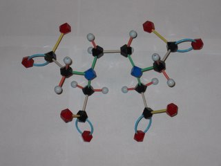
Orac has been posting about the abuse of chelation therapy for treating autism and other disorders. So what's a chelate and how does it work to remove metal ions from the body? EDTA is shorthand for ethylenediamminetetraacetic acid, which has the structure shown at the left. The disodium calcium salt of EDTA is the usual chemotheraputic form. Lone pairs of electrons on the nitrogens and oxygens of the EDTA (tagged blue and red in the photo) latch onto the metal. This Lewis acid-base reaction results in the metal being sequestered inside the EDTA molecule. Tucked away inside the EDTA, the metal can't accumulate in the body's tissues and is eventually eliminated. EDTA has different affinities for different metal ions, but is a pretty effective scavenger of most metal ions, including iron and calcium. Removal of too much calcium can result in cardiac arrest, so EDTA is not without safety isses, as Orac points out!

The word chelation come from the Greek for claw. Molecules that attach to metals at multiple points, like EDTA, are called multidentate ligands from their capacity to "bite" onto the metal. EDTA makes a hexadentate metal-ligand complex (6 points of attachment) with some ions, a pentadentate complex with others.

Weird Words of Science 8: Ligands, the ties that bind


Many transition metals react with bases (such as ammonia) to produce beautifully colored transition metal-ligand complexes. The word ligand comes from the Latin ligare which means to tie or bind. The same root leads to ligaments, which tie your bones together.

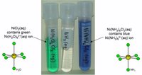
The photo shows green Ni(H2O)62+ and blue green Ni(NH3)62+. The ligands are water and ammonia respectively, "tied" to the Ni(II) center. The ligands form an octahedron around the metal center.

Trojan Horse Molecules: Penicillin


Penicillin was one of the first antibiotics in wide use. It was discovered in the late 19th century by a French medical student (Ernest Duchesne), though his work was never pursued. Fleming independently discovered the antibacterial activity of Penicillium mold derivatives in 1928. The active molecule was difficult to extract. The compound was finally synthesized in 1957 by John Sheehan, a chemist at MIT. This feat was made possible by the determination of penicillin's structure in 1944 by Dorothy Crowfoot Hodgkin, an X-ray crystallographer who won the 1964 Nobel prize in chemistry for that discovery and many others (including B-12 and insulin).

How does penicillin work? It is a Trojan horse molecule. Penicillin disrupts the synthesis of bacterial cell walls, thus inhibiting the bacteria's reproduction. The enzyme responsible for assembling the cell walls picks up penicillin, thinking it can incorporate into the wall. Unfortunately for the bacteria, the penicillin molecule opens up and destroys the enzyme's ability to function.

The key step in this sneak attack is the nucleophilic attack of the enzyme onto an electrophilic site on the four-membered β-lactam ring. We've been discussing these reactions in my general chemistry class this week.


Watch this webcast if you want to see how the reaction works and learn a bit about nucelophilic reactions.

Elemental Tales: Get the lead out!

Workers manufacturing the pigment white lead (Pb(OH)2.2PbCO3 apparently made a habit of adding dilute sulfuric acid to their drinking water to prevent lead poisoning. The reaction of the sulfate ions (SO42-) with the aqueous lead ions (Pb2+) forms an insoluble precipitate of lead sulfate, effectively removing the lead from the water (as long as you let the precipipate settle before drinking!). This risk of lead poisoning in these workers was so high that it was referred to as "painter's colic".

van Gogh's Palette

In an attempt to brighten a dreary Philadelphia day, I pulled out a coffee mug that glows with Vincent van Gogh's sunflowers. Among the most vivid of his favorite pigments is chrome yellow. Chrome yellow was first isolated from a natural source (the mineral crocoite) in the late 18th century by Parisienne chemist Vauquelin. By the late 19th century, when van Gogh's sunflowers took form, the vibrant yellow was one of a series of new and exceptionally vivid colors. Chrome yellow is actually a lead salt, lead chromate (PbCrO4. The pigment isstill used today but it has been replaced in many cases by similarly colored, less toxic organic pigments. Unfortunately chrome yellow degrades over time, so that the once brilliantly glowing sunflowers now appear to be dry, drab ocher shadows of van Gogh's vision.

Perhaps influenced by the mug, this week's webcast general chemistry example problem is based on a simple inorganic synthesis of the chrome yellow pigment. One of my colleague's uses another synthesis. in her course on "The Stuff of Art"


Read more about the history and chemistry of color in Bright Earth: Art and the Invention of Color by Philip Ball.

Making a Mark

My interest in MRI has become less academic. I need an MRI of my hand. The orthopedic surgeon noted in passing that they will mark the spot of interest with a capsule of vitamin E, in the same way that they use lead markers in X-rays. I wondered what was so special about the vitamin E that left a trace in the MRI. Turns out that the spin-lattice relaxation time (T1) of the H's in tocopherol's chain of -CH2s is very short, and provides a high intensity signal which can be used to mark the spot. Mineral oil will work, too, but the vitamin E capsules are convenient.

A Magnetic Moment

The Culture of Chemistry welcomes 2006 - now that the grading is done and vacation has begun for me in earnest.

Graham at "Over My Med Body" notes that the total radiation dose in a year from natural background sources is much larger than the dose from any single test. He notes that ultrasound and MRIs are exceptions: ultrasound uses sound waves, and MRIs use magnets. What exactly do those magnets do?

The nuclei of many atoms have "spin" states. Like quarks which have a property called by physicists "color" but are not actually different colors like socks, spin is an instrinsic property of nuclei but this does not necessarily mean that the atoms are spinning like the earth! Hydrogen atoms, of which there are many in the human body (more than 10 pounds worth) have two spin states. Not every atom has multiple spin states. Carbon-12 (the most common form of carbon) has only one spin state. So what happens in an MRI? Radiation (yes, radiation, just very, very low energy radiation) in the form of radio waves forces the hydrogen nuclei to change state to the higher energy spin state. The time it takes for the hydrogens to relax to their low energy spin state is measured. There are two ways for the hydrogen atom to "lose spin", one is called spin-lattice relaxation (T1), the other is spin-spin relaxation (T2). Hydrogen atoms in different environments relax at different rates. Hydrogens in fatty tissue, for example, have very different relaxation times than watery tissue.

So if the changes happen because of radiation, what are the magnets for? It turns out that the separation between spin states depends on the magnitude of the magnet field, as well as the magnetic moment of the nucleus. In the earth's field, the energy between spin states is too small to do the trick of exciting them up to the higher energy state and watching them fall down. You need a high magnet field to do this.